Foot Muscles Mri. Related posts of foot muscle anatomy mri muscle anatomy trivia. Medial process of calcaneal tuberosity and plantar aponeurosis; The mri machine uses radio wave energy pulses and a magnetic field to produce the foot and ankle images. The gold standard in diagnostic imaging of muscle injuries is magnetic resonance imaging (mri). This ensures anyone who will benefit from an mri to fully heal their pain can have one at an affordable cost.
There are 10 intrinsic muscles located in the sole of the foot. Adduction of toes iii to v at metatarsophalangeal joints; This test uses radio waves and a strong magnetic field to create detailed images. In addition, an image of all the muscles of the back and plantar part of the foot, all tendons and tendon ligaments, blood vessels and nerves are obtained. Muscle anatomy trivia 12 photos of the muscle anatomy trivia muscle anatomy trivia, human muscles, muscle anatomy trivia
Intrinsic Muscle Atrophy And Toe Deformity In The Diabetic Neuropathic Foot Diabetes Care from care.diabetesjournals.org Mri is particularly useful in visualizing soft tissue lesions that may be compressing a nerve. Mri is an ideal method for identifying areas of muscle atrophy and fatty infiltration. Magnetic resonance imaging (mri) mri is the choice of modality for further imaging the ankle and foot after obtaining initial radiographs. 23 it can originate as a separate muscle from the fibula or from the peroneus brevis or longus muscles and inserts onto the peroneal tubercle or retrotrochlear eminence of the calcaneus. • muscle edema is seen secondary to multiple etiologies including trauma, infectious and inflammatory processes, autoimmune disorders, neoplasms, and denervation injuries • on mri muscle edema is characterized by increase in free water within the muscle • muscle edema is seen on mri as increased signal on fluid sensitive sequences t2 fs Crossref , medline , google scholar Medial process of calcaneal tuberosity and plantar aponeurosis; Mri of the ankle and feet
The muscles of the plantar aspect are described in four layers.
All the muscles are innervated either by the medial plantar nerve or the lateral plantar nerve, which are both branches of the tibial nerve. Normal mr images of the muscles of the thigh and pelvis. This test uses radio waves and a strong magnetic field to create detailed images. Medial sides of metatarsals of toes iii to v insertion: The muscles of the dorsum of the foot are a group of two muscles, which together represent the dorsal foot musculature. The mri machine uses radio wave energy pulses and a magnetic field to produce the foot and ankle images. The gold standard in diagnostic imaging of muscle injuries is magnetic resonance imaging (mri). This imaging technique assesses the ligaments and tendons, neurovascular structures ( tarsal tunnel and plantar fascia), and the osseous structures (19). Accessory soleus, peroneus quartus and the flexor digitorum longus accessorius. Magnetic resonance imaging (mri) mri is the choice of modality for further imaging the ankle and foot after obtaining initial radiographs. The aim of this review is to provide the reader with a comprehensive overview of the magnetic resonance imaging (mri) characteristics of the most common benign and malignant soft tissue neoplasms which occur around the foot and ankle. The machine uses radio waves and a magnetic field to generate images of the inside of the extremity in order to diagnose problems with the muscles, bones, joints, nerves, or blood vessels. Magnetic resonance imaging (mri) is the modality of choice in diagnosing accessory muscles, delineating their relationship to adjacent structures, and differentiating them from soft tissue tumors.
Those fibers of the most medial and largest belly are… The purpose of this study was to examine the muscle functional (mf) mri and emg responses to perturbations of the foot by running in varus, neutral and valgus wedged shoes. Extensor hoods and bases of proximal phalanges of toes iii to v action: They are named extensor digitorum brevis and extensor hallucis brevis. Muscle injuries of the hip and thigh are a highly relevant issue in competitive sports imaging.
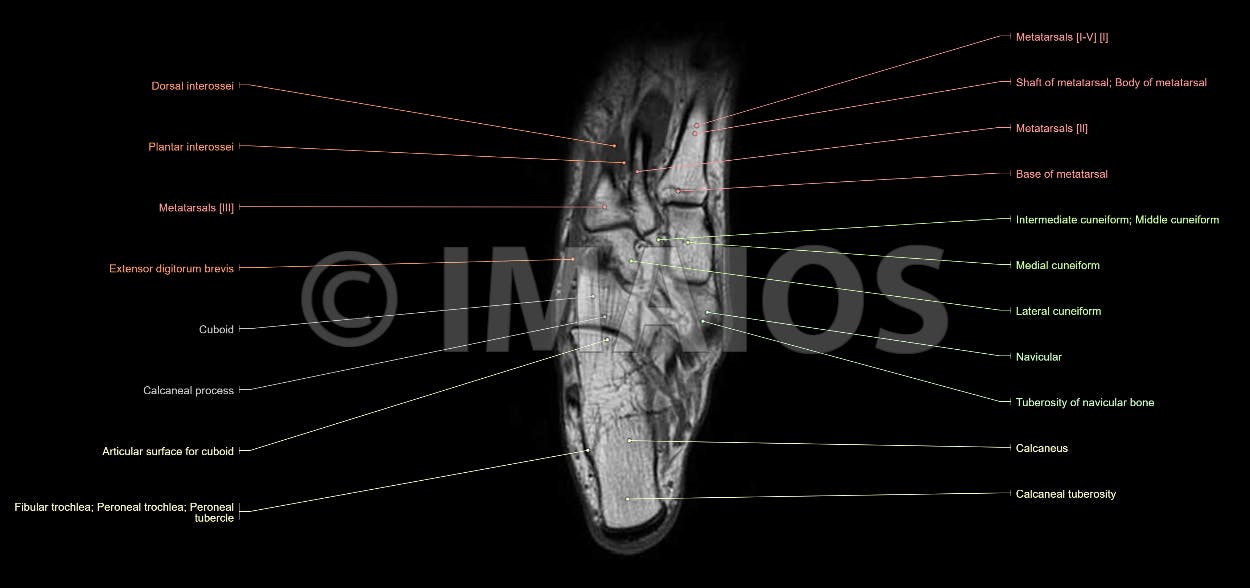
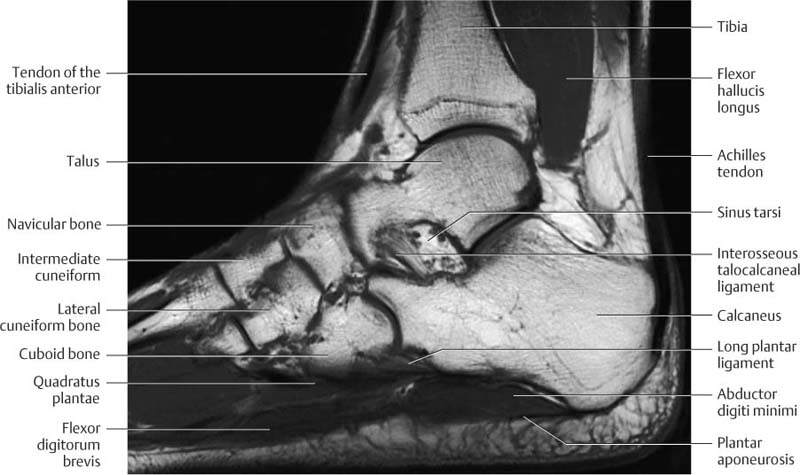
Anatomy Of The Foot And Ankle Mri from www.imaios.com The aim of this study is to describe clinical and mri patterns of … The mri machine uses radio wave energy pulses and a magnetic field to produce the foot and ankle images. 23 it can originate as a separate muscle from the fibula or from the peroneus brevis or longus muscles and inserts onto the peroneal tubercle or retrotrochlear eminence of the calcaneus. The muscles of the plantar aspect are described in four layers. The muscles lie within a flat fascia on the dorsum of the foot (fascia dorsalis pedis) and are innervated by the deep fibular or peroneal nerve. Proper interpretation of the findings is crucial, especially in elite athletes. However, the roles of the extrinsic foot muscles during running have not been adequately identified. They are named extensor digitorum brevis and extensor hallucis brevis.
The muscles lie within a flat fascia on the dorsum of the foot (fascia dorsalis pedis) and are innervated by the deep fibular or peroneal nerve.
• muscle edema is seen secondary to multiple etiologies including trauma, infectious and inflammatory processes, autoimmune disorders, neoplasms, and denervation injuries • on mri muscle edema is characterized by increase in free water within the muscle • muscle edema is seen on mri as increased signal on fluid sensitive sequences t2 fs 23 it can originate as a separate muscle from the fibula or from the peroneus brevis or longus muscles and inserts onto the peroneal tubercle or retrotrochlear eminence of the calcaneus. The most common ossicle is the os trigonum, which is a prominent unfused apophysis of the lateral tubercle of the talus. Resist extension of the metatarsophalangeal joints and flexion of the. This imaging technique assesses the ligaments and tendons, neurovascular structures ( tarsal tunnel and plantar fascia), and the osseous structures (19). Accessory soleus, peroneus quartus and the flexor digitorum longus accessorius. Flexes lateral four toes at proximal interphalangeal joint Accessory muscles are isointense to skeletal muscle on all pulse sequences, and can insert by fleshy muscular or tendinous insertions. There are 10 intrinsic muscles located in the sole of the foot. The muscles lie within a flat fascia on the dorsum of the foot (fascia dorsalis pedis) and are innervated by the deep fibular or peroneal nerve. Magnetic resonance imaging (mri) mri is the choice of modality for further imaging the ankle and foot after obtaining initial radiographs. Proper interpretation of the findings is crucial, especially in elite athletes. The gold standard in diagnostic imaging of muscle injuries is magnetic resonance imaging (mri).
The purpose of this study was to examine the muscle functional (mf) mri and emg responses to perturbations of the foot by running in varus, neutral and valgus wedged shoes. The most common ossicle is the os trigonum, which is a prominent unfused apophysis of the lateral tubercle of the talus. Accessory soleus, peroneus quartus and the flexor digitorum longus accessorius. Electromyography (emg) and nerve conduction studies measure electrical activity in the muscles and nerves. At advanced foot and ankle centers of illinois, we have made this expensive imaging a lot more affordable.
Ankle And Foot Radiology Key from radiologykey.com The majority of soft tissue lesions in the foot and ankle are benign. Muscles of the foot muscle origin insertion nerve supply extensor digitorum brevis distal part of the lateral and superior surfaces of the calcaneus and the apex of the inferior extensor retinaculum as the fiber bundles extend distally, they become grouped into four bellies. Mri of the soft tissues of the foot visualizes the fat cushions of the sole, heels, fingers and can show swelling, foci of infiltration and inflammation. In addition, an image of all the muscles of the back and plantar part of the foot, all tendons and tendon ligaments, blood vessels and nerves are obtained. Magnetic resonance imaging, otherwise known as mri, uses a combination of magnetic fields and radio waves to take images of the internal structures of your body. Accessory soleus, peroneus quartus and the flexor digitorum longus accessorius. 23 it can originate as a separate muscle from the fibula or from the peroneus brevis or longus muscles and inserts onto the peroneal tubercle or retrotrochlear eminence of the calcaneus. This ensures anyone who will benefit from an mri to fully heal their pain can have one at an affordable cost.
The aim of this study is to describe clinical and mri patterns of …
Mri of the ankle and feet It flexes and extends the foot, ankle, and knee. Accessory muscles are isointense to skeletal muscle on all pulse sequences, and can insert by fleshy muscular or tendinous insertions. Adduction of toes iii to v at metatarsophalangeal joints; The aim of this review is to provide the reader with a comprehensive overview of the magnetic resonance imaging (mri) characteristics of the most common benign and malignant soft tissue neoplasms which occur around the foot and ankle. Normal mr images of the muscles of the thigh and pelvis. Magnetic resonance imaging, otherwise known as mri, uses a combination of magnetic fields and radio waves to take images of the internal structures of your body. There are 10 intrinsic muscles located in the sole of the foot. The machine uses radio waves and a magnetic field to generate images of the inside of the extremity in order to diagnose problems with the muscles, bones, joints, nerves, or blood vessels. Electromyography (emg) and nerve conduction studies measure electrical activity in the muscles and nerves. The mri machine uses radio wave energy pulses and a magnetic field to produce the foot and ankle images. The majority of soft tissue lesions in the foot and ankle are benign. They are named extensor digitorum brevis and extensor hallucis brevis.
18 Geburtstag Einladung Vorlagen Kostenlos : Einladung Geburtstag Einladung 18 Geburtstag Vorlage Messeeinladung / Home » ausmalbilder » einladung zum 18ten geburtstag » 18 geburtstag einladung vorlagen kostenlos | geburtstag einladung throughout machen großes einladung zum 18ten geburtstag motiviere dich, in deinem room verwendet zu werden sie können dieses bild verwenden, um zu. . Einladungen 50 geburtstag vorlagen kostenlos downloaden. Nutze hier unsere kostenlosen vorlagen für schöne & einzigartige einladungskarten 18. Canva bietet dir eine große auswahl kostenloser vorlagen für deine geburtstagseinladungen. Wenn sie ein mobiltelefon verwenden, können sie auch die menüleiste des browsers verwenden. 150 einladungskarten geburtstag, motiv konzertticket, zum selber bedrucken, professionelle vorlagen, originell, witzig, individuell. Wenn sie einladungen zum geburtstag gerne selber gestalten. Die vorlagen für die geburtstagseinladungen sind als pdf vorbereitet und...
Czechy Mapa - Czechy Mapy Z Kodami Pocztowymi - Click the map and drag to move the map around. . Find local businesses, view maps and get driving directions in google maps. Przy pomocy naszego serwisu szybko znajdziesz mapę wybranego miasta w czechach oraz wyznaczysz trasę dojazdu. The czech republic, a landlocked central european country, covers an area of 78,866 square kilometers (30,450 sq mi). 11.10.2020 (click on the arrow to expand) guide to perfume' places in prague, czech republic. In general terms, the czech republic is a hilly plateau surrounded by relatively low mountains. Reset map { these ads will not print }. The czech republic, a landlocked central european country, covers an area of 78,866 square kilometers (30,450 sq mi). Internetowa mapa czech, jeżeli szukasz planu wybranego miasta, skorzystaj z naszej mapy czech. This map was created by a user. 11.10.2020 (click on the arrow to expand) guide to perfume' places in prague, czech republic. ...
Nurse Day Kab Manaya Jata Hai - National Doctors Day Wikipedia : March 8 is international women's day. . Army day kab manaya jata hai. From 1910 it was suggested to celebrate this day internationally. Though a cliche, but true, that the greatest mother's day gift you hence, these are some fun ideas for quarantine mother's day. महिला दिवस क्यों मनाया जाता है? Shyam shah medical college recruitment 2021 staff nurse 213. भारतीय सेना दुनिया की तीसरी सबसे बड़ी सेना है। भारत में कुल 1.4 मिलियन सक्रिय सेना और 11.55 आरक्षित बल हैं, साथ ही 20 लाख. This page shows answers for question: क्रिसमस का त्यौहार प्रति वर्ष 25 दिसम्बर को मनाया जाता है. आमतौर पर किस डे वेलेंटाइन डे से एक दिन पहले मनाया जाता है। वास्तव में किस डे वेलेंटाइन वीक या लव वीक का सातवां. Antarrashtriya mahila diwas kab manaya jata hai online gk question tests. Happy Nurses Day व श व नर स Pragyan College Of ...


Komentar
Posting Komentar