Upper Leg Tendon Anatomy / Upper leg muscles, artwork - Stock Image - F005/5442 ... : The largest muscle masses in the leg are present in the thigh and the calf.
Dapatkan link
Facebook
X
Pinterest
Email
Aplikasi Lainnya
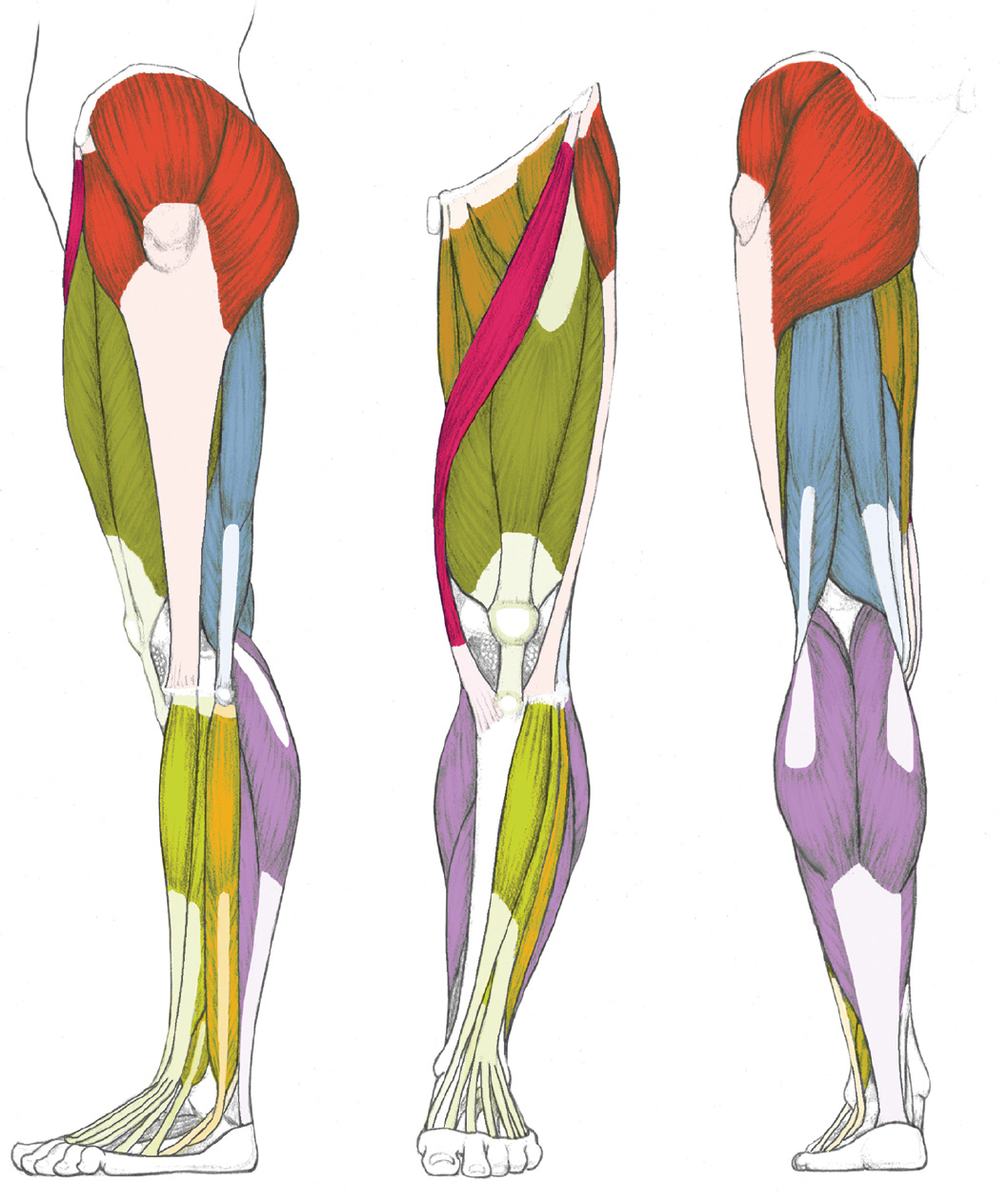
Upper Leg Tendon Anatomy / Upper leg muscles, artwork - Stock Image - F005/5442 ... : The largest muscle masses in the leg are present in the thigh and the calf.. Upper leg tendon anatomy : The four muscles all extend the lower leg. We study anatomy at the practical anatomy class we study the human body. It serves to attach the plantaris, gastrocnemius (calf) and soleus muscles to the calcaneus (heel) bone. The thigh muscles are divided into three compartments:
•medial thigh muscles•adductor longus muscle•adductor magnus muscle•adductor. Muscles that move the hip and thigh. The upper leg is composed of the femur the hamstring tendon is also connected to the tibia, immediately below the rear of the knee joint. Other muscles of the anterior (front) thigh include the pectineus, sartorius,. Related posts of muscle anatomy upper leg.
LEFT: Lateral view from schoolbag.info Its muscle belly is on the back aspect of the upper arm. Muscles in the anterior compartment of the thigh. Upper leg tendon anatomy : The knee joint is the junction of the thigh and leg. This is why you have to indicate which biceps you are taking about when discussing one or other of these muscles. This important tendon in the back of the calf and ankle stores the elastic energy needed for running, jumping, and other physical activity. It is also visible on the medial edge of the thigh from the anterior. Your upper leg includes seven major muscles.
It serves to attach the plantaris, gastrocnemius (calf) and soleus muscles to the calcaneus (heel) bone.
Learn about the muscles, tendons, bones, and ligaments that comprise the knee joint anatomy. Tendons of the lower leg, muscles tendons and ligaments of the upper leg. It serves to attach the plantaris, gastrocnemius (calf) and soleus muscles to the calcaneus (heel) bone. Possibly the most important tendon in terms of mobility is the achilles tendon. This long muscle extends from the pelvis to the. The long head originates from the ischial tuberosity of the pelvis. Tendons are cords made of tough tissue, and they work as special connector pieces between bone and muscle. Muscles in the anterior compartment of the thigh. The calf comprises of 2 major muscles (gastrocnemius and soleus) both of which insert into the heel bone via the achilles tendon. Upper leg tendon anatomy from i0.wp.com the achilles tendon or heel cord, also known as the calcaneal tendon, is a tendon at the back of the lower leg, and is the. This is the group of muscles that you often see body builders flexing, which protrude just above the knee and take up most of the upper leg. Tendons are also bands of connective tissue. Tendons are thick bands of tissue that connect muscles to bone.
It is also visible on the medial edge of the thigh from the anterior. It's the area that runs from the hip to the knee in each leg. This long muscle extends from the pelvis to the. Muscles that move the hip and thigh. Tendons are also bands of connective tissue.
Upper Leg Tendon Anatomy - 3d Human Upper Leg Anatomy Or ... from thumbs.dreamstime.com It's the area that runs from the hip to the knee in each leg. It runs straight down the leg and attaches to the patella via the quadriceps femoris tendon. Lateral (fibular) collateral ligament (fcl) upper part middle part lower part popliteus tendon (pt) upper part i. Ebraheim's educational animated video describes muscle anatomy of the thigh. The four muscles all extend the lower leg. We study anatomy at the practical anatomy class we study the human body. Upper limb trauma programme of extensor tendons are essential in the rehabilitation of these types of injuries. Muscles in the anterior compartment of the thigh.
Learn about the muscles, tendons, bones, and ligaments that comprise the knee joint anatomy.
Your lower leg includes three main muscles, located behind your tibia or shinbone. Squeeze your knees together and boom, you're contracting the adductors. It's the area that runs from the hip to the knee in each leg. The hamstrings are three muscles at the back of the thigh that affect hip and knee movement. The human leg, in the general word sense, is the entire lower limb of the human body, including the foot, thigh and even the hip or gluteal region. Putra bagus mei 16, 2021. Meanwhile, the vastus lateralis is on the side of the thigh, while the vastus intermedius is hidden below the rectus femoris(5). Rectus femoris these four muscles come together to form a single tendon, which inserts into the patella, or kneecap. People who play soccer have these specific muscles of the leg very well defined, so they're like a walking anatomy atlas for thigh muscles. •medial thigh muscles•adductor longus muscle•adductor magnus muscle•adductor. This mri wrist coronal cross sectional anatomy tool is absolutely free to use. The thigh is the region between the hip and knee joints. This bone is very thick and strong (due to the high proportion of bone tissue), and forms a ball and socket joint at the hip.
Its muscle belly is on the back aspect of the upper arm. Notice the upper leg has a biceps muscle just like the upper arm does. On the medial edge of the posterior thigh is the gracilis muscle. We study anatomy at the practical anatomy class we study the human body. The knee joint is the junction of the thigh and leg.
Related Pictures anterior and posterior views of the human ... from i.pinimg.com On the medial edge of the posterior thigh is the gracilis muscle. The rectus femoris is located in the center of the thigh, while the vastus medialis is in the middle of the said body part. The short head originates from the linea aspera on posterior surface of the femur. The four muscles all extend the lower leg. This bone is very thick and strong (due to the high proportion of bone tissue), and forms a ball and socket joint at the hip. Putra bagus mei 16, 2021. Upper leg anatomy and function the upper leg is often called the thigh. Rectus femoris these four muscles come together to form a single tendon, which inserts into the patella, or kneecap.
The upper leg is composed of the femur the hamstring tendon is also connected to the tibia, immediately below the rear of the knee joint.
Your upper leg includes seven major muscles. Possibly the most important tendon in terms of mobility is the achilles tendon. Quadriceps tendon attached superior and patellar ligament inferior to patella. Meanwhile, the vastus lateralis is on the side of the thigh, while the vastus intermedius is hidden below the rectus femoris(5). Search photos retinaculum from t3.ftcdn.net 3d illustration back fit strong human anatomy. The largest muscle masses in the leg are present in the thigh and the calf. Upper leg tendon anatomy : Because the hamstrings cross the back of the hip joint on their way to the knee, they help to extend the hip. Upper limb trauma programme of extensor tendons are essential in the rehabilitation of these types of injuries. Squeeze your knees together and boom, you're contracting the adductors. Tendons are also bands of connective tissue. The knee joint is the junction of the thigh and leg. •medial thigh muscles•adductor longus muscle•adductor magnus muscle•adductor.
18 Geburtstag Einladung Vorlagen Kostenlos : Einladung Geburtstag Einladung 18 Geburtstag Vorlage Messeeinladung / Home » ausmalbilder » einladung zum 18ten geburtstag » 18 geburtstag einladung vorlagen kostenlos | geburtstag einladung throughout machen großes einladung zum 18ten geburtstag motiviere dich, in deinem room verwendet zu werden sie können dieses bild verwenden, um zu. . Einladungen 50 geburtstag vorlagen kostenlos downloaden. Nutze hier unsere kostenlosen vorlagen für schöne & einzigartige einladungskarten 18. Canva bietet dir eine große auswahl kostenloser vorlagen für deine geburtstagseinladungen. Wenn sie ein mobiltelefon verwenden, können sie auch die menüleiste des browsers verwenden. 150 einladungskarten geburtstag, motiv konzertticket, zum selber bedrucken, professionelle vorlagen, originell, witzig, individuell. Wenn sie einladungen zum geburtstag gerne selber gestalten. Die vorlagen für die geburtstagseinladungen sind als pdf vorbereitet und...
Czechy Mapa - Czechy Mapy Z Kodami Pocztowymi - Click the map and drag to move the map around. . Find local businesses, view maps and get driving directions in google maps. Przy pomocy naszego serwisu szybko znajdziesz mapę wybranego miasta w czechach oraz wyznaczysz trasę dojazdu. The czech republic, a landlocked central european country, covers an area of 78,866 square kilometers (30,450 sq mi). 11.10.2020 (click on the arrow to expand) guide to perfume' places in prague, czech republic. In general terms, the czech republic is a hilly plateau surrounded by relatively low mountains. Reset map { these ads will not print }. The czech republic, a landlocked central european country, covers an area of 78,866 square kilometers (30,450 sq mi). Internetowa mapa czech, jeżeli szukasz planu wybranego miasta, skorzystaj z naszej mapy czech. This map was created by a user. 11.10.2020 (click on the arrow to expand) guide to perfume' places in prague, czech republic. ...
Nurse Day Kab Manaya Jata Hai - National Doctors Day Wikipedia : March 8 is international women's day. . Army day kab manaya jata hai. From 1910 it was suggested to celebrate this day internationally. Though a cliche, but true, that the greatest mother's day gift you hence, these are some fun ideas for quarantine mother's day. महिला दिवस क्यों मनाया जाता है? Shyam shah medical college recruitment 2021 staff nurse 213. भारतीय सेना दुनिया की तीसरी सबसे बड़ी सेना है। भारत में कुल 1.4 मिलियन सक्रिय सेना और 11.55 आरक्षित बल हैं, साथ ही 20 लाख. This page shows answers for question: क्रिसमस का त्यौहार प्रति वर्ष 25 दिसम्बर को मनाया जाता है. आमतौर पर किस डे वेलेंटाइन डे से एक दिन पहले मनाया जाता है। वास्तव में किस डे वेलेंटाइन वीक या लव वीक का सातवां. Antarrashtriya mahila diwas kab manaya jata hai online gk question tests. Happy Nurses Day व श व नर स Pragyan College Of ...


Komentar
Posting Komentar